
These effects can conveniently be modeled at a mesoscopic spatial scale with electric dipoles distributed along the cortical mantle (green arrows in figure below). Mass effects of close-to-simultaneous changes in post-synaptic potentials across the cell group add up in time and space. The reason is essentially in the morphology and mass effect of these cells, which present elongated shapes, and are grouped in large assemblies of cells oriented in a similar manner approximately normal to the cortex. Overall, it is assumed that most of - but not exclusively - the MEG/EEG signals are generated by postsynaptic activity of ensembles of cortical pyramidal neurons of the cerebral cortex. Note that, as always with modeling, we need to deal with various degrees of approximation. To better understand how forward and inverse modeling work, we need to have a basic understanding of the physiological origins of MEG/EEG signals. To sort out this question will influence the time and computational resources required for data analysis (source analysis multiplies the needs in terms of disk storage, RAM and CPU performance).

Nevertheless, if your neuroscience question can be solved by measuring signal latencies over broad regions, or other aspects which do not depend crucially on anatomical localization (such as global signal properties integrated over all or clusters of sensors), source modeling is not required. This is not an issue with EEG, with essentially one sensor type (electrodes, dry or active, all measuring Volts). This problem does not exist in EEG, where electrodes are attached to the head and arranged according to standard positions.Īnother important point to consider when interpreting MEG sensor maps and that can be solved by working in the MEG source space instead, is that MEG manufacturers use different types of sensor technology (e.g., magnetometers vs. Therefore, between two runs of acquisition, or between subjects with different head shapes and sizes and positions under the helmet, the same MEG sensors may pick up signals from different parts of the brain. Hence data sensor topographies depend on the position of the subject's head inside the MEG sensor array. Indeed in MEG and contrarily to EEG, the head of the participant is not fixed with respect to sensor locations. In MEG, source maps can be a great asset to alleviate some issues that are specific to the modality. Moving to the source space can help discriminating between contributing brain regions. In EEG in particular, scalp topographies are very smooth and it is common that contributions from distant brain regions overlap over large clusters of electrodes. Source mapping is a form of spatial deconvolution of sensor data.
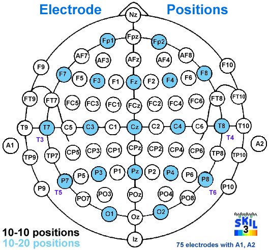
EEG SENSOR REGISTRATION
after averaging across multiple participants) is limited by the accuracy of anatomical registration of individual brain structures, which are very variable between participants, and intersubject variations in functional specialization with respect to cortical anatomy. As for other imaging modalities, spatial resolution of group-level effects (i.e. The spatial resolution of MEG and EEG depends on source depth, the principal orientation of the neural current flow, and overall SNR: still, a sub-centimeter localization accuracy can be expected in ideal conditions, especially when contrasting source maps between conditions in the same participant. Source estimation improves anatomical resolution further from the interpretation of sensor patterns. Empirical interpretations of sensor topographies can inform where brain generators might be located: which hemisphere, what broad aspect of the anatomy (e.g., right vs. If one of your primary objectives is to identify and map the regions of the brain involved in a specific stimulus response or behavior, source estimation can help address this aspect. Although Brainstorm simplifies the procedures, it is important to decide whether source modeling is essential to answer the neuroscience question which brought you to collect data in the first place. Reconstructing the activity of the brain from MEG or EEG recordings involves several sophisticated steps.


 0 kommentar(er)
